Recovering a fossil can take longer than the discovery announcement suggests. In its 16 November 2026 notice marking ninety years of research at Sterkfontein, Wits University announced recognition for fourteen fossil technicians. Their work included finding bones, freeing them from rock, cleaning fragments and making casts through which other people could study them. The anniversary brings a practical question into view: how does material inside a South African cave become evidence about human origins? The history of Little Foot, a nearly complete Australopithecus skeleton, shows why the answer involves patient observation and several different kinds of knowledge. [1]
Sterkfontein lies northwest of Johannesburg. Wits’ December 2018 account describes a recovery that began with re-examination of stored material. In 1994 and 1997, Ronald Clarke recognised foot and lower-leg bones that had been classified among animal remains. Finding these fragments did not expose an entire skeleton. Clarke, Stephen Motsumi and Nkwane Molefe had to locate the matching breaks in bones still embedded underground. The stored pieces and the cave deposits were connected through an anatomical fit: evidence in a collection directed a search back to a specific place. [2]
The same account distinguishes fourteen years of excavation from six years of cleaning and reconstruction. Hammer and chisel removed overlying material; an air scribe, a vibrating needle-like tool, helped expose bone more delicately. These stages matter because a skeleton is also a record of what happened after death. Wits describes displacement, crushing and later mineral deposits. A fracture need not record an injury during life, and material surrounding a bone need not have formed at the same time. Interpreting the specimen therefore depends on preserving relationships between bone, broken surfaces and sediment during recovery. [2]
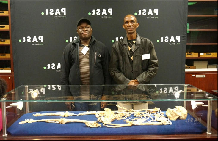
A March 2026 commentary in the South African Journal of Science explains the judgement involved in preparation. A technician anticipates which anatomical structure may continue inside hard, cemented sediment, consulting comparative bones and casts rather than treating every obstruction as expendable rock. The authors describe Abel Molepolle’s work with a compressor-driven air scribe and his later collaboration with Andrew Phaswana on cleaning and casting. Their archive includes a December 2017 photograph of the two technicians beside the original Little Foot skeleton, reproduced here as historical evidence of that work, rather than an image of the anniversary. [3]
That history continues through people learning the work. In an undated profile published by the Sterkfontein research site, Itumeleng Shadrack Molefe recalls visiting the caves during school holidays while his father Nkwane worked there. He later sought employment in gold mining before joining the Sterkfontein team in 2013. His responsibilities include identifying, cleaning, sorting and cataloguing fossils. The account describes an occupation combining excavation with knowledge of anatomy and geology, where a find has to be recognised before it can become a research specimen. Familiarity with a place is a beginning; the profile also documents acquired professional skill. [4]
Molefe’s ambitions include learning more about casting from his cousin Molepolle. That detail makes the transmission of expertise concrete: observing bones, learning preparation and producing replicas are related but distinguishable abilities. His recollection also resists a convenient heroic ending. Years may pass without a major discovery, while identification and cataloguing continue. A scientific collection grows through this repeated work, not only through exceptional finds. His own account expresses both attachment to Sterkfontein and a desire to improve his skills; neither can be reduced to an anonymous supporting role in somebody else’s discovery. [4]
Preserving a bone is only part of preserving evidence. A 2015 PaleoAnthropology paper on digitising the Little Foot excavation describes the problem of recording a skeleton distributed through an irregular, sloping deposit before its removal. The researchers used laser scanners to create a three-dimensional surface record. A laser stripe is projected onto a surface and observed by a camera at a known position; the distorted line allows distances to be calculated. Repeated views can then be combined. This is surface measurement, distinct from the X-ray techniques later used to examine structures within the skull. [5]
The team reported difficulty positioning scanners in the cave, shadows and gaps where surfaces were hidden from view. Some colour information was unusable, while processing took much longer than acquisition. A digital reconstruction consequently involves choices about joining measurements, reducing detail and filling gaps. The paper acknowledges Motsumi and Molefe’s help with digitisation. Its contribution is a record of spatial relationships that could no longer be inspected in precisely the same arrangement after excavation. A model preserves access to part of a setting; it does not eliminate the work of interpreting that setting. [5]
A different view became possible when Little Foot’s skull was scanned at Diamond Light Source in Britain in 2019. Amélie Beaudet and colleagues’ 2021 eLife paper reports a synchrotron X-ray investigation of dental and cranial tissues. Rather than physically cutting sections through the specimen, the team reconstructed internal structures from scans. They imaged the whole skull and selected regions at finer resolutions, reaching a voxel size of 3.25 micrometres for some observations. A voxel is a small three-dimensional unit of the reconstructed image: the scale describes the recorded volume, rather than a guarantee that every structure is equally visible. [6]
The images revealed fine tooth tissues and channels within bone. The researchers also observed enamel defects consistent with disruptions during development. Such traces can support questions about an individual’s life without identifying a unique cause: illness and nutritional stress are among the possibilities discussed. This preliminary study examined one fossil. The distinction between seeing a feature and explaining its origin remains essential. Higher resolution makes particular structures available for study; it does not, by itself, settle their biological meaning. [6]
In a paper published on 2 March 2026 in Comptes Rendus Palevol, Beaudet and colleagues used the scan data to reconstruct Little Foot’s damaged face. Bone and teeth were digitally separated from sediment; fragments were repositioned according to anatomical correspondences, and an intact portion on one side helped reconstruct the other. The researchers compared facial measurements and configurations of thirty-four anatomical landmarks with other fossils and living primates. This made a distorted specimen available to comparisons that its buried condition had obstructed. The reconstruction was a documented analytical operation, rather than a photograph of the face as it looked during life. [7]
Their analysis found closer facial-shape similarities with the Ethiopian Australopithecus specimen A.L. 444-2 than with Sts 5 from Sterkfontein. The authors nevertheless identify limits: the fossil sample is small, variation within groups is incompletely known, and their reconstruction could be refined. Differences between sexes and uncertainty about classification also affect interpretation. Similarity does not establish a direct ancestral relationship or a particular migration route. The study opens possibilities about relationships across eastern and southern Africa while leaving alternatives to be tested. [7]
Beaudet and Dominic Stratford’s own March 2026 account of this research explains why access to the interior required substantial collaboration. Dense mineral infill had limited conventional scanning. The skull travelled from South Africa to Diamond in 2019, and processing the resulting image stacks required major computing resources. Their account connects curation, transport, imaging and reconstruction instead of presenting a digital face as an isolated technological achievement. The geographical journey of the specimen is part of the method: the fossil remained the basis of the study, while different facilities supplied complementary capacities to examine it. [8]
The same authors distinguish a reconstruction from an evolutionary explanation. Facial form can bear on feeding, vision and interaction with an environment, but several mechanisms may contribute to anatomical change. Their account presents proposed relationships between fossils as possibilities constrained by a limited comparative sample. Digital access therefore extends a question rather than bringing inquiry to an automatic conclusion. For readers encountering a striking reconstructed image, that distinction is useful: the image makes damaged anatomy intelligible, while the argument still depends on the comparisons chosen and on what the surviving material can support. [8]
The anniversary notice also records how long preparation occupies a working life. Wits reports that Molepolle spent years in the grotto and then years in the laboratory, followed by the making of moulds and casts. The recognition announced for fourteen technicians includes retired colleagues and people honoured posthumously. It recognises a history extending beyond one specimen, and reminds readers that the production of usable evidence depends on sustained work across generations. [1]
There is a documented omission to correct here, without claiming that every earlier account erased technicians. In March 2026, the South African Journal of Science’s editorial accompanying the tribute explicitly acknowledged that the journal had overlooked important contributors who might not qualify as authors under existing guidelines. It invited discussion of hidden contributions to research. The editors also cautioned against turning recognition into paternalistic celebration. That is a more precise problem than a general accusation against science: a publication can acknowledge a person yet still leave readers unable to understand the knowledge that person contributed. [9]
Reading the excavation, imaging and technicians’ accounts together makes that contribution visible through practice. The journal’s editorial asks for histories of how research is made, including work that publication conventions can obscure. Little Foot offers such a history: recognising a fragment, retaining its context, preparing a surface and comparing a reconstruction each make different claims possible. Recognition is strongest when it explains those relationships, rather than adding names to an unchanged discovery story. The anniversary is an opportunity to present African scientific work through the skills that produce evidence and the care with which conclusions remain open to revision. [9]
- [1]Wits University. Sterkfontein celebrates 90 years by honouring the technicians behind its discoveries2026-11-16
- [2]Wits University. Little Foot’s history revealed for the first time2018-12-20
- [3]South African Journal of Science; Clarke and colleagues. Tribute to Abel Molepolle and the central role of the Sterkfontein Caves technicians in international palaeoanthropological research2026-03-26
- [4]Wits Sterkfontein Caves. Itumeleng Shadrack Molefe: Can Little Foot be topped?Undated; accessed 2026-11-19
- [5]PaleoAnthropology; Subsol and colleagues. In Situ 3D Digitization of the “Little Foot”2015; accepted 2015-03-01
- [6]eLife; Beaudet and colleagues. Preliminary paleohistological observations of the StW 573 (“Little Foot”) skull2021-03-02; scans 2019
- [7]Comptes Rendus Palevol; Beaudet and colleagues. Virtual reconstruction and comparative study of the face of StW 573 (“Little Foot”)2026-03-02; scans 2019
- [8]The Leakey Foundation; Amélie Beaudet and Dominic Stratford. A new face for “Little Foot”, the most complete Australopithecus skeleton to date2026-03-11; author account republished from The Conversation
- [9]South African Journal of Science. Making the hidden visible: A challenge and call for submissions2026-03/04; accessed 2026-11-19
Did this publication help you understand the topic?
0 voluntary responses. Browser-based participation; responses are not a representative survey.
We welcome suggestions, contributions and recommendations.
Editorial standards ↗